3D Templates of Brain Development – First Steps in Building a Resource Bank for the Study of Brain Developmental Phases
The IBEN Atlas project, 3D templates for brain development, has led to the creation of detailed maps showing the structure and organization of brain regions at different developmental stages from embryonic stage E15 to 8 days after birth in mice models. These atlases help researchers and physicians better understand the localization of brain areas involved in various functions and study neurological diseases.
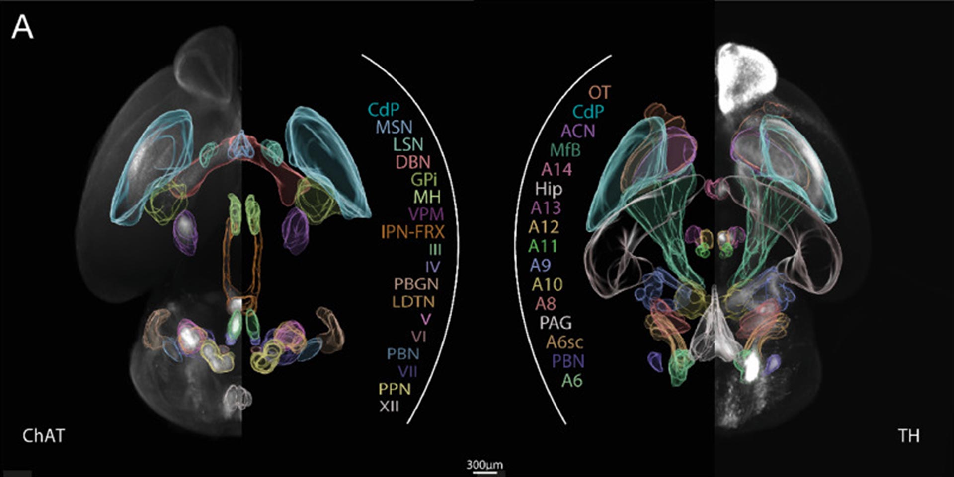
We are proud of our contribution to scientific research by co-publishing the very first 3D atlases of cholinergic, dopaminergic, and noradrenergic cells for the developing brain.
The Context
From the embryonic stage to birth, a remarkable sequence of events unfolds, shaping the complex development of a living organism at various scales. From the intrauterine stages to the perinatal and postnatal phases, the organism, and especially the brain, undergoes a symphony of genesis, maturation, migration, connection, and growth, orchestrating the miracle of life. These events leave an indelible imprint at multiple levels, from the subtleties of the transcriptome and cells to the structural intricacies of the connectome. Each step of this fascinating process contributes to the foundation of our existence.
However, this developmental period is extremely sensitive, and numerous disruptors, some of which are still unknown, can alter this magnificent machinery. From the transcriptome to the connectome, including biological and cognitive functions, each level can be affected by these external and/or genetic influences.
Our Goal
The objective of this project is to design quantitative 3D atlases, in other words, 3D brain maps, of specific neuronal populations, from the early stages of pregnancy to birth and beyond, in mice models.
Our method
To achieve this, B&A Biomedical has leveraged its expertise in analyzing images obtained from advanced microscopic acquisition (Light Sheet). This technique enables the optical slicing of whole rodent brain samples that have been rendered transparent using the clearing technique (iDISCO). The neuronal populations of interest are labeled with fluorescent antibodies. These multiple samples are then treated using various image processing techniques such as registration methods, allowing for precise alignment of different brain structures according to developmental stages and fluorescence channels.
Results
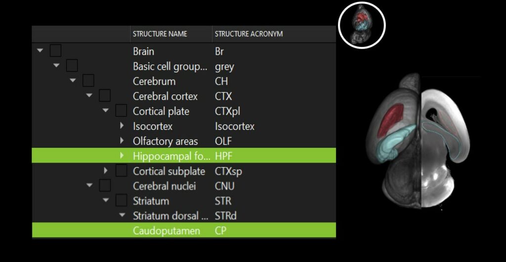
We have been able to generate templates that account for interindividual morphological variations, each one representing an average brain and its internal structures at a given developmental stage. Our 3D atlases provide essential comparative references for studying structural differences between physiological and pathological development. They serve as valuable tools for the scientific community, paving the way for groundbreaking discoveries on the physiological and pathological processes of brain development.
Download the IBEN ATLAS
We strongly believe in the importance of sharing knowledge and fostering collaboration in the fields of health and biology. That is why we have decided to make the IBEN Atlas: developmental brain atlases from embryonic stage E15 to 8 days after birth in mice models, freely accessible and available for download (download links below).
In return, we strongly encourage you to cite our publication when using our resources in your research, publications, or presentations. Your citation will not only acknowledge our contribution but will also help us continue to provide high-quality resources to the scientific community.
The user instructions and Python source codes are freely available on our GitHub repository.
From this project, we also recognized the urgent need for user-friendly tools for complex image analysis, accessible to biology researchers without extensive programming or mathematical expertise. We observed that manual processing methods are time-consuming, require significant human and computational resources, and often lead to results that are difficult to reproduce. In response to this challenge, we committed to developing innovative algorithms inspired by our experience and expertise in image analysis, including registration, segmentation, and cell counting. Our goal is to empower researchers by providing intuitive and efficient solutions, enabling them to unleash their scientific creativity and fully explore the vast potential of imaging data in healthcare and biology.
Contact Us
If you are interested in our expertise and the scope of our services, or if you would like to share an idea for a collaborative project, please feel free to contact us :
To do so, kindly use the form below. We will respond as quickly as possible. (All fields are mandatory)
